
Imaging Department

Overview
Imaging diagnosis (Radiology) is a medical specialty that applies modern imaging techniques such as X-ray, Ultrasound, Computed Tomography (CT), Magnetic Resonance Imaging (MRI) and Nuclear Medicine to support diagnosis, monitoring and treatment of diseases. These techniques allow detailed investigation of anatomical morphology and structure, as well as assessment of certain organ functions in the body, helping to early detect abnormalities related to pathology; thereby supporting treatment planning or directly participating in interventional treatment.
Today, imaging diagnosis plays an indispensable role in all areas of health care: from screening, detection and early-stage disease diagnosis; assessing disease severity and progression; to treatment prognosis. In particular, minimally invasive image-guided interventional procedures increasingly contribute positively to treatment, making patients safer, recovering faster and achieving higher effectiveness.
Alongside the explosion of science and technology, imaging equipment is continually improved in hardware design, image processing software, automation and artificial intelligence integration, contributing to increased diagnostic accuracy, shortened examination time, and optimized safety for patients.
In addition to modern equipment, the department's doctors and technicians constantly maintain a spirit of learning, research and daily knowledge updates. Since its establishment together with the University of Medicine and Pharmacy Hospital in Ho Chi Minh City in 1994, the Imaging Department has continuously developed, now capable of receiving and examining from 5,000 to over 6,000 patients each day, becoming one of the largest and most robustly specialized units in the country.
With the aim of optimizing patient experience, the Imaging Department provides multiple result delivery options via text messages, the UMC Care app, or direct printed reports. In particular, X-ray, ultrasound, CT, and MRI image data of inpatients or outpatients are stored long‑term on a modern PACS system for easy retrieval, comparison of lesion progression, and to provide the most comprehensive diagnosis.
Unit
Original Form for Vascular Intervention
Strength
The motto of the Department of Imaging Diagnostics at Ho Chi Minh City University of Medicine and Pharmacy Hospital: “Accurate in-depth diagnosis – Maximally effective intervention – Modern – Collaborative – Trustworthy”. Our department has built a deep specialization model, gathering doctors rigorously trained in specific fields such as neurology, respiratory, cardiovascular, breast, digestive–hepatobiliary, obstetrics‑gynecology, musculoskeletal, vascular and interventional radiology. Consequently, the department plays a core role in multidisciplinary case conferences, providing accurate, specialized and personalized imaging for each patient, optimally supporting multimodal treatment strategies and modern personalized medicine.
Through daily scheduled multidisciplinary specialty conferences by subspecialty, doctors regularly cross‑reference imaging with clinical data, pathology and treatment outcomes. This process enables our imaging physicians to continuously build internal expertise, develop deep experience and sustainably improve their skills.
About Ultrasound:
Performs most types of ultrasound such as general ultrasound, vascular Doppler ultrasound, obstetric‑gynecologic ultrasound, software‑assisted ultrasound, musculoskeletal ultrasound,…
Performs specialized ultrasounds such as: intra‑operative ultrasound, transplant ultrasound, trans‑rectal prostate ultrasound, especially endorectal and transrectal ultrasound. The department is the first in the country to introduce this technique for diagnosing anorectal diseases such as anal fistula, perianal abscess, colorectal cancer, anal cancer,…

About Neuro‑cranial Imaging:
The department has full imaging equipment for diagnosing neurological conditions: multi‑slice CT scanner, 1.5T and 3T MRI systems used for routine and advanced imaging.
The department can perform all basic and advanced neuro‑MRI techniques thanks to strong collaboration with internal and external clinical neurologists.
Stroke group: In collaboration with emergency physicians and neurologists, perform rapid and effective imaging surveys of ischemic and hemorrhagic stroke. Combined with modern equipment and a team of neurologic experts, can conduct multi‑territory CTA, perfusion CT to assess vessels, infarct core, penumbra for appropriate intervention; the 1.5T and 3T MRI systems can perform MRA, MRP sequences, and high‑resolution vessel wall imaging (HR‑MRI). Notably, the department has implemented RAPID software for rapid diagnosis, planning, and early treatment of stroke patients.
Neurosurgery group: Performs advanced techniques such as DTI, fMRI for pre‑operative mapping and functional assessment, intra‑operative navigation and postoperative prognosis. MRP, MRS for evaluating brain lesions, inflammation, tumors, distinguishing residual tumor from postoperative necrosis. DTI assesses invasive nerve fiber tracts of brain tumors pre‑operatively; CSF‑flow imaging evaluates cerebrospinal fluid dynamics, flow rate, …
Endovascular intervention group: Can perform real‑time dynamic imaging sequences (TWIST) especially for assessing cerebrovascular malformations.
Movement disorder group: Performs techniques such as high‑resolution MRI to assess the “owl’s eye” sign, DTI, MRS, fMRI for evaluating neurological and cognitive function, … MRI diagnosis of Parkinson’s disease with high‑resolution SWI sequences.
About Head‑Neck Imaging:
Diagnose diseases of oral‑maxillofacial, ENT, ophthalmology, oncology, …
Diagnose and biopsy pulmonary and mediastinal diseases. Notably, the department is the first unit to perform MRI of mediastinal masses to enhance diagnostic accuracy of mediastinal tumors.
About Cardiac‑Vascular Imaging:
Using CT, MRI with dedicated software, the department has performed:
Coronary artery imaging techniques, evaluation of congenital heart diseases, cardiomyopathies, valvular diseases,…
Complex vascular imaging techniques for diagnosing pulmonary artery and aortic diseases, meeting needs for surgical or endovascular treatment planning, monitoring, and post‑treatment assessment.
About Digestive, Hepatobiliary, Urogenital Imaging:
This is one of the traditional strengths of the Department of Imaging Diagnostics.
Along with the development of the hospital’s digestive, hepatobiliary, and transplant surgery departments, the department has performed specialized techniques for accurate diagnosis of the following disease categories:
Liver: Evaluate liver tumors using MRI, CT, MRI with liver‑specific contrast agents, assess pre‑ and post‑liver transplantation for donors and recipients, monitor progression of cirrhosis, hepatitis, liver tumors before and after treatment.Biliary‑pancreatic system: Diagnose all biliary, gallbladder, and pancreatic diseases such as inflammation, stones, various tumors,…
Digestive tract: Perform specialized techniques to evaluate disorders such as dysphagia, constipation, esophageal, gastric, colorectal, and anal cancers, gastrointestinal bleeding diseases; CT or MRI enterography to assess small bowel conditions such as tumors, tuberculous enteritis, Crohn’s disease, inflammatory bowel disease, chronic intestinal obstruction, CT virtual colonoscopy. Notably, the department is the first in the country to implement dynamic MRI to assess pelvic floor compartments in conditions like constipation, defecatory disorders, urinary incontinence, uterine prolapse, pelvic floor prolapse,…
Urogenital: Besides diagnosing common conditions such as stones and urinary trauma, the department performs imaging to evaluate pre‑ and post‑kidney transplantation, and assesses prostate diseases with dynamic MRI,…
Abdominal cavity: Assess intra‑abdominal abscesses in postoperative complications, intraperitoneal and retroperitoneal masses.
About Obstetric‑Gynecologic Imaging:
This remains one of the leading strengths of the Department of Imaging Diagnostics in using MRI to evaluate uterine pathology: anomalies, intra‑myometrial masses, cervical tumors, endometrial diseases,… It also assesses adnexal pathology including masses, abscesses, tubo‑ovarian disease, fetal anomalies,…
About Musculoskeletal Imaging:
Early detection, accurate diagnosis, pre‑operative planning and post‑operative monitoring of joint diseases such as cartilage‑bone grafts, post‑surgical complications, prosthetic joint fractures,…
Early detection, accurate diagnosis and post‑treatment monitoring of intra‑articular diseases such as ankylosing spondylitis, rheumatoid arthritis,… helping patients avoid or limit disabling complications.
CT scanner with high contrast and high‑resolution multiplanar capability to assess thin ligament, tendon, and cartilage structures, features to reduce artifact from surgical implants, shorten scan time, lower radiation dose, and cover large joint areas.
X‑ray and CT of body joints in all necessary positions (static, weight‑bearing).
About Interventions:
Perform extracorporeal interventions such as fine‑needle aspiration (FNA), biopsy of various tumors, high‑frequency ablation, microwave ablation, vacuum‑assisted suction of benign breast tumors, drainage of abscesses or fluid collections,…
Training & Research
The Department of Imaging Diagnostics is a training base for imaging physicians and imaging technicians. Therefore, during its operations, the department regularly organizes continuous education programs (CME), short‑term training courses, and technology transfer activities to enhance the skills of other healthcare units.
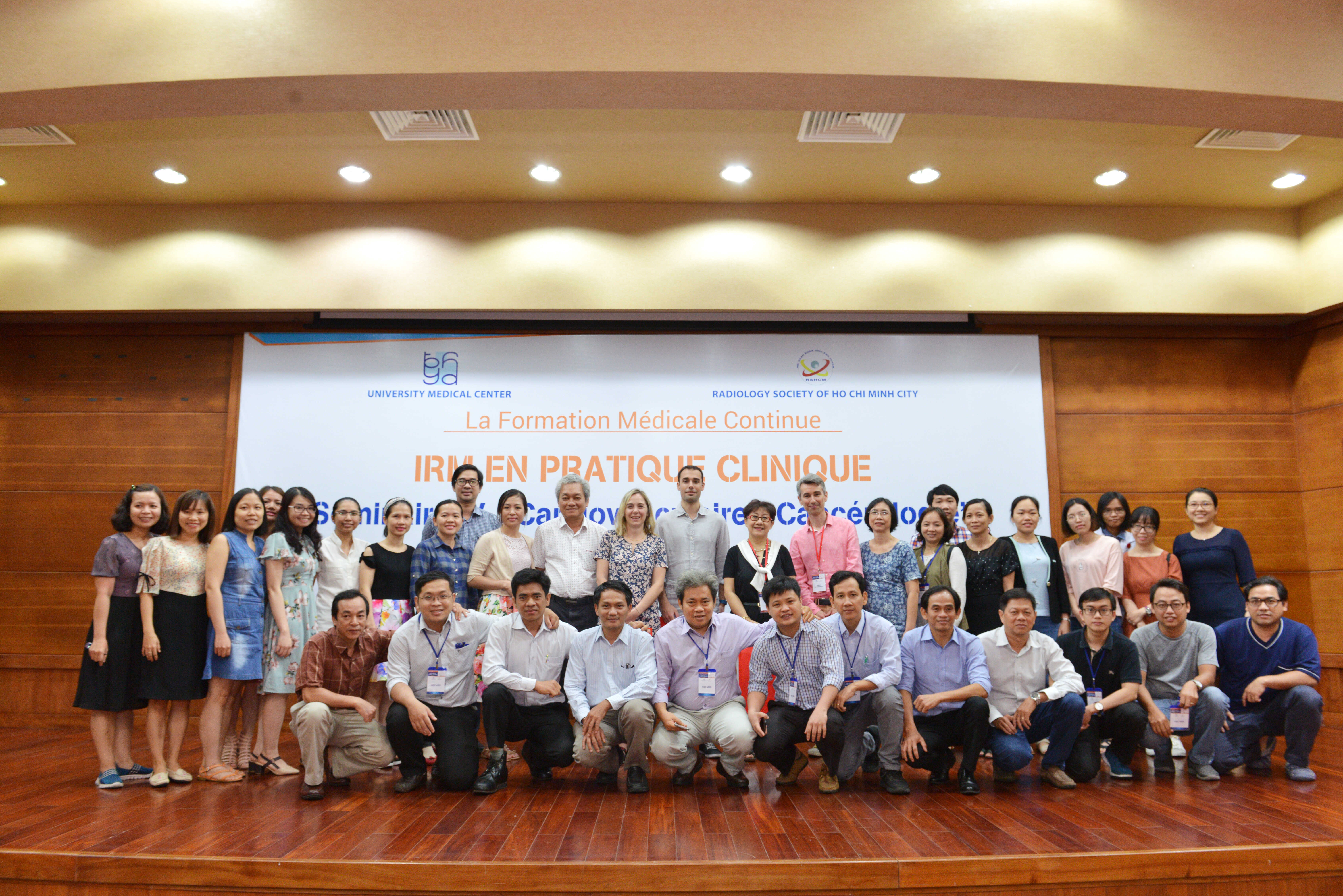
Khoa Chẩn đoán hình ảnh thực hiện đào tạo - hợp tác trong môi trường quốc tế để mở rộng chuyên môn: Hợp tác đào tạo với các Viện - Trường trên thế giới; Cử nhân viên đi học tập ở Mỹ, Anh, Pháp, Singapore, Hàn Quốc, Đài Loan, Thái Lan; Nhận học viên từ các Viện – Trường trên thế giới đến tham quan học tập; Tổ chức các hội thảo, hội nghị, khóa huấn luyện chuyên môn và mời các chuyên gia nước ngoài đến giảng dạy, thực hành.
Trong lĩnh vực Siêu âm: Khoa Chẩn đoán hình ảnh là nơi đào tạo siêu âm căn bản và nâng cao cho các bác sĩ sau đại học lâu năm của miền Nam. Trung bình mỗi năm khoa đào tạo hơn 100 học viên với các trình độ cơ bản, nâng cao và bổ túc sau đại học. Song song đó, Khoa tham gia nghiên cứu nhiều đề tài về siêu âm, tổ chức hội thảo và viết sách siêu âm atlas cho học viên, thường xuyên cập nhật kiến thức mới trong lĩnh vực siêu âm trên thế giới.
Trong lĩnh vực can thiệp: Khoa nhận đào tạo các lớp can thiệp nội và ngoại mạch cho các bác sĩ sau đại học của các tỉnh thành phía Nam. Đồng thời chuyển giao các kỹ thuật can thiệp nội và ngoại mạch cho các bệnh viện trong khu vực.
Khoa là nguồn cung cấp dữ liệu thực hiện đề tài nghiên cứu khoa học cho học viên sau đại học, nội trú, cao học, chuyên khoa 2, tiến sĩ với hơn 100 bài báo công bố trong nước và quốc tế.
Đồng chủ nhiệm đề tài nhánh 1 đề tài nghiên cứu của Bộ Khoa học Công nghệ
Chủ nhiệm và tham gia hơn 5 đề tài nghiên cứu khoa học của Sở Khoa học Công nghệ.
Với đội ngũ bác sĩ giàu kinh nghiệm, say mê nghiên cứu, giỏi ngoại ngữ, trong thời gian qua, khoa đã có hơn 20 bài báo quốc tế đăng trên các tạp chí ISI/ Scopus với vai trò tác giả và đồng tác giả trong nhiều lĩnh vực chuyên ngành.
Equipments
Carestream's PACS system, with full modern features, has connected all imaging systems of X‑ray, Ultrasound, CT, MRI within the hospital as well as external (partner) facilities to transmit, store data and process images, enabling film‑free operation, health‑insurance billing, and integration with the hospital’s electronic medical records.
The ultrasound system includes nearly 30 modern devices from brands such as GE, Siemens, Samsung, BK, Esaote, etc. Numerous advanced software are equipped beyond classic B‑mode ultrasound, such as Doppler ultrasound, multi‑point elastography, 3D endorectal and transrectal ultrasound, Biplane prostate ultrasound, Navigation positioning system, intra‑operative ultrasound, …
In addition, the department is equipped with a variety of probes to serve various specialties. Alongside, essential modern equipment (operated under ultrasound guidance) such as multi‑needle high‑frequency tumor ablation devices, microwave tumor ablation devices, vacuum‑assisted breast tumor suction devices, … are present. All this modern machinery and equipment are available in more than 25 advanced ultrasound rooms of the department to deliver accurate results with images stored and managed on the common system and PACS.
X‑ray: 5 digital high‑frequency systems, 1 digital fluoroscopy system, 1 high‑end breast imaging system, 4 digital mobile X‑ray units, 1 panoramic dental imaging system and 1 cone‑beam dental imaging system, all connected to PACS.
CT: Techniques performed on three systems including 64‑slice, 128‑slice and >256‑slice CT scanners (Definition AS and AS Plus – Siemens; Revolution – GE) with dedicated software for cardiovascular, neuro, thoracic, musculoskeletal, digestive, hepatobiliary, …
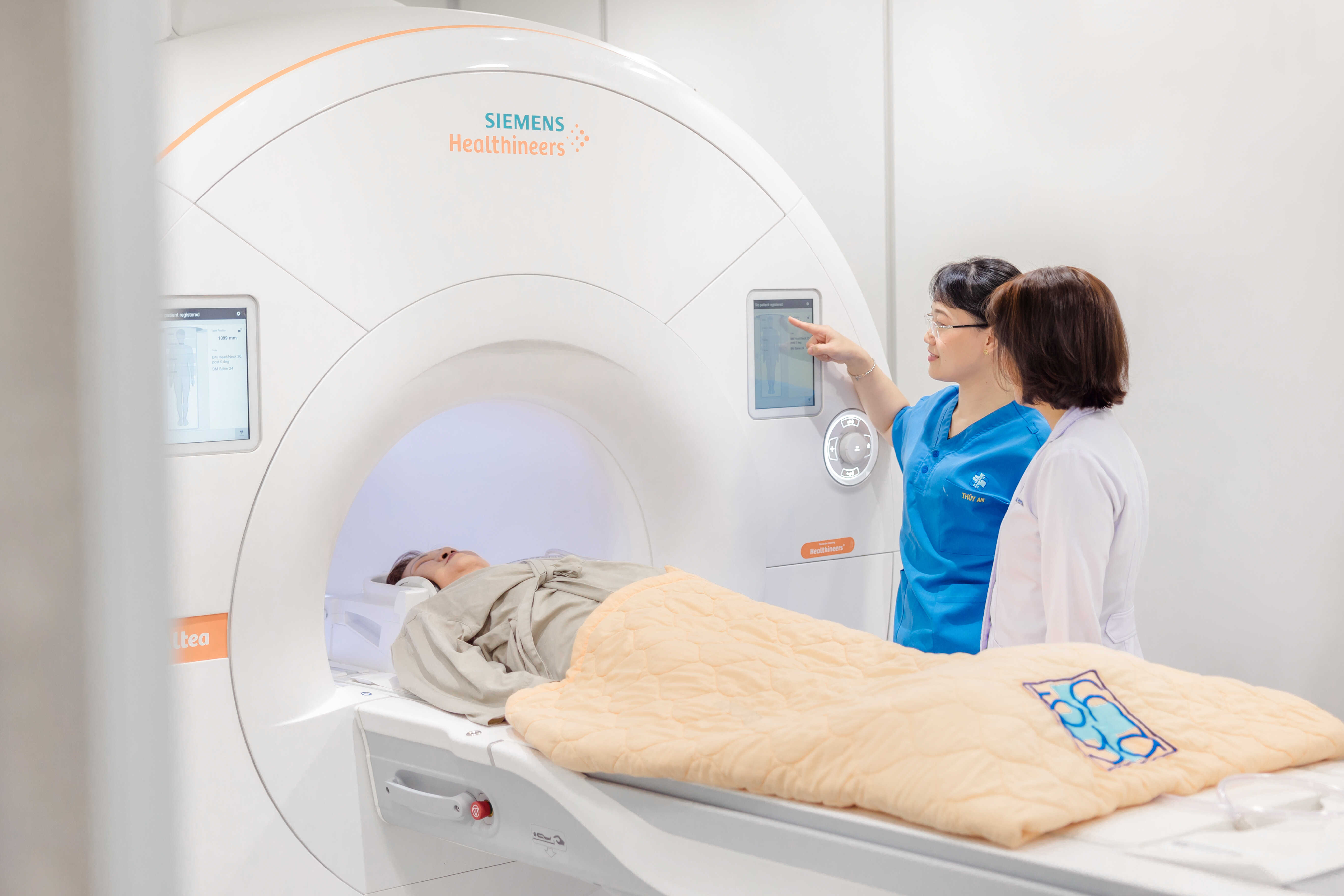
MRI: Techniques performed on four systems comprising two 1.5‑Tesla units (Avanto – Siemens, Altea – Siemens) and two 3‑Tesla units (Verio – Siemens, Lumina – Siemens).


Staff
As a Department of a leading University Hospital nationwide, alongside the mission of diagnosis and treatment, we also serve as a major training facility for undergraduate and postgraduate levels; training the department’s personnel to be highly qualified, systematic and scientific. Most department staff hold postgraduate or university degrees:
- 02 Associate Professors
- 05 Doctor physician
- 11 Doctor physician Second‑level Specialist
- 60 Masters and first‑level specialists
- 32 Bachelor's in Imaging Technology and Nursing