

Breast biopsy: From atypical hyperplasia to cancer
Breast biopsy is an important procedure for diagnosing breast abnormalities. However, many women feel confused by the complex medical terminology used in their pathology reports. This article provides a detailed explanation of atypical hyperplasia, carcinoma in situ (including ductal carcinoma in situ [DCIS] and lobular carcinoma in situ [LCIS]), and invasive breast carcinoma. It aims to help you understand the meaning of each diagnosis and its clinical significance so that you can make informed decisions about appropriate treatment.
1. Key takeaways
- Breast biopsy is essential for accurately diagnosing breast lesions [1].
- Atypical hyperplasia (AH) is a precancerous condition that increases the risk of developing breast cancer in the future [1, 3].
- Carcinoma in situ (CIS, including DCIS and LCIS) refers to non-invasive cancer or a high-risk lesion confined to the milk ducts or lobules [1, 3, 8].
- Invasive breast carcinoma occurs when cancer has spread beyond the milk ducts or lobules into surrounding tissue and has the potential to metastasize [4].
- Understanding these terms helps patients and doctors make appropriate decisions about treatment and follow-up [1, 3].
2. Atypical hyperplasia (AH)
Definition
Atypical hyperplasia (AH) is an abnormal proliferation of epithelial cells in the breast that does not meet the criteria for a diagnosis of carcinoma in situ [1]. It is considered a precancerous condition or a high-risk lesion and is often detected incidentally during a breast biopsy [1, 3].
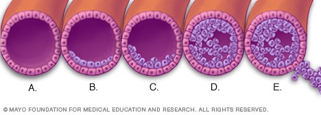
Classification
AH is divided into two main types:
- Atypical ductal hyperplasia (ADH): Abnormal cell proliferation within the milk ducts [1, 3].
- Atypical lobular hyperplasia (ALH): Abnormal cell proliferation within the breast lobules [1].
Management and follow-up
- For ADH diagnosed through core needle biopsy, surgical excision of the lesion is generally recommended to rule out underlying DCIS or invasive cancer that may not have been detected in the initial sample [1, 3, 5]. The rate of upgrade to cancer following a core needle biopsy diagnosis of AH ranges from 10% to 30% [1, 4].
- For ALH, surgical excision is more controversial. Selected patients may instead undergo close surveillance or receive medical therapy, such as tamoxifen, to reduce their risk [1, 3].
- Both conditions require enhanced screening and close follow-up, such as more frequent mammography, potentially combined with magnetic resonance imaging (MRI) [1, 3].
3. Carcinoma in situ (CIS, including DCIS and LCIS)
3.1. General definition
Carcinoma in situ refers to the presence of cancer cells or high-risk abnormal cells in the breast that have not invaded through the basement membrane into surrounding tissues [1, 8]. It includes ductal carcinoma in situ (DCIS) and lobular carcinoma in situ (LCIS) [1, 8].
Ductal carcinoma in situ (DCIS)
- Definition: DCIS occurs when cancer cells are present within the breast milk ducts but have not invaded through the basement membrane into surrounding tissues [1, 9]. It is a non-invasive form of breast cancer [1].
- Histological characteristics and classification: DCIS is classified as low-, intermediate-, or high-grade based on cellular and architectural features [3, 9]. High-grade DCIS is often associated with comedo-type necrosis [3].
- Clinical significance and cancer risk: DCIS has the potential to progress to invasive breast cancer if left untreated [1, 9]. There is also a significant risk of local recurrence or the development of invasive cancer in the same breast [1].
- Diagnosis and treatment: Diagnosis is primarily established through breast biopsy following the detection of abnormalities on imaging, such as microcalcifications on mammography [1, 3, 9]. Treatment generally involves breast-conserving surgery (lumpectomy) or mastectomy, with radiation therapy considered depending on tumor characteristics and other risk factors [1, 9].
Lobular carcinoma in situ (LCIS)
- Definition: LCIS is characterized by the proliferation of abnormal cells within the breast lobules and terminal duct-lobular units [8]. Despite the word “carcinoma” in its name, LCIS is NOT considered a true cancer. Instead, it is a marker of increased risk for developing invasive breast cancer [8].
- Histological characteristics: Small, uniform, discohesive cells, resulting from loss of E-cadherin expression, fill and expand the breast lobules [8].
- Clinical significance and cancer risk: LCIS increases the risk of developing invasive breast cancer in both breasts, with an approximately 7- to 12-fold increase compared with the general population [8]. This risk applies to any type of invasive breast cancer, whether ductal or lobular, rather than only lobular cancer [8].
- Management and follow-up: LCIS generally does not require extensive surgical excision. Management typically involves close surveillance, including enhanced screening with mammography and MRI. Preventive medication, such as tamoxifen or raloxifene, may also be considered to reduce breast cancer risk [8].
4. Invasive breast carcinoma
Definition
Invasive breast cancer occurs when cancer cells break through the basement membrane of the milk ducts or lobules and spread into the surrounding breast tissue [1]. These cancer cells can metastasize to other parts of the body through the lymphatic system or bloodstream [1].
Main types
- Invasive ductal carcinoma (IDC): This is the most common type of invasive breast cancer, accounting for approximately 70–80% of all cases [4, 11]. Cancer cells originate in the milk ducts and break through the duct walls to invade the surrounding breast tissue [4].
- Invasive lobular carcinoma (ILC): This is the second most common type, accounting for approximately 10–15% of invasive breast cancer cases [1, 10]. It originates in the milk-producing lobules and tends to spread in a different pattern, making it more difficult to detect on imaging [1].
Clinical features and diagnosis
Invasive breast cancer can present with various signs and symptoms, including a palpable lump in the breast or armpit, breast swelling, changes in the size or shape of one breast, skin dimpling or an orange-peel appearance, nipple changes such as inversion, redness, or scaling, and abnormal nipple discharge [1].
Diagnosis is confirmed through core needle biopsy or open surgical biopsy, together with imaging tests such as mammography, ultrasound, and magnetic resonance imaging (MRI) [1, 12].
Clinical significance and treatment
The treatment plan for invasive breast cancer depends on several factors, including the stage of the disease, cancer type, hormone receptor status (estrogen receptor [ER] and progesterone receptor [PR]), HER2 status, and the patient's overall health [1, 10]. The main treatment options include:
- Surgery: Lumpectomy or mastectomy, often accompanied by axillary lymph node surgery [1, 10].
- Radiation therapy: Uses high-energy X-rays to destroy any remaining cancer cells after surgery or to shrink tumors [1, 10].
- Chemotherapy: Uses medications to destroy cancer cells and may be given before surgery (neoadjuvant therapy) or afterward (adjuvant therapy) [1, 10].
- Hormone therapy: Used for hormone receptor-positive tumors to block the hormones that stimulate cancer cell growth [1, 10].
- Targeted therapy: Uses medications that target specific characteristics of cancer cells, such as HER2 positivity [1, 10].
5. When should you see a doctor?
You should seek medical attention promptly if you notice any unusual breast changes, including:
- Any new lump in the breast or armpit [1].
- Changes in the size or shape of one breast [1].
- Breast skin dimpling, puckering, a rough texture, or redness [1].
- Nipple inversion, changes in nipple shape, scaling, or abnormal discharge [1].
- Persistent breast pain with no clear cause [1].
In addition, anyone who has received an abnormal breast biopsy result, such as a diagnosis of atypical hyperplasia or carcinoma in situ, should consult a specialist for an assessment and guidance on the clinical significance of the findings, as well as an appropriate follow-up or treatment plan [1, 13]. Women with a family history of breast cancer or other risk factors should consult their doctors about appropriate screening and effective risk management [1].
6. Frequently asked questions (FAQ)
- Is atypical hyperplasia cancer? Atypical hyperplasia (AH) is not cancer. It is a precancerous condition in which abnormal cell proliferation increases the risk of developing breast cancer in the future [1].
- If I am diagnosed with atypical hyperplasia, will I need surgery? For atypical ductal hyperplasia (ADH) diagnosed through core needle biopsy, surgical excision is generally recommended to ensure that no underlying invasive cancer or DCIS has been missed [1, 3]. For atypical lobular hyperplasia (ALH), the decision to perform surgery is less clear-cut, and management often involves surveillance or preventive therapy [1].
- Is carcinoma in situ, including DCIS and LCIS, dangerous? DCIS is a form of non-invasive cancer that may progress to invasive cancer if left untreated. LCIS is not cancer but indicates an increased risk of developing invasive breast cancer in either breast in the future [1, 8]. Both conditions require appropriate management and close follow-up.
- What is the main difference between DCIS, LCIS, and invasive breast cancer? DCIS consists of cancer cells confined to the milk ducts, while LCIS consists of abnormal cells confined to the lobules. Neither has invaded through the basement membrane [1, 8]. Invasive breast cancer occurs when cancer cells break through the basement membrane and spread into surrounding tissues, with the potential to metastasize to other parts of the body [1, 10].
- What can I do to reduce my breast cancer risk after being diagnosed with atypical hyperplasia or carcinoma in situ? Measures include close medical follow-up, lifestyle changes such as eating a healthy diet, exercising regularly, maintaining a healthy weight, and limiting alcohol consumption. Preventive medication, such as tamoxifen, may also be considered under medical supervision [1, 8].
- Is breast biopsy painful, and is it safe? Breast biopsy is usually performed under local anesthesia, so patients generally do not feel pain during the procedure. It is considered a safe procedure with a low complication rate. The most common complications are bruising or mild bleeding at the biopsy site [12].
7. References
- [1] Myers, D. J., & Walls, A. L. (2023). Atypical Breast Hyperplasia. StatPearls - NCBI Bookshelf. Accessed: 2025-12-11.
- [2] Mayo Clinic. Atypical hyperplasia of the breast - Symptoms and causes. Accessed: 2025-12-11.
- [3] Tomlinson-Hansen, S. E., Khan, M., & Cassaro, S. (2023). Atypical Ductal Hyperplasia. StatPearls - NCBI Bookshelf. Accessed: 2025-12-11.
- [4] Hodorowicz-Zaniewska, D., Brzuszkiewicz, K., Szpor, J., Kibil, W., Matyja, A., Dyląg-Trojanowska, K., Richter, P., & Szczepanik, A. M. (2018). Clinical predictors of malignancy in patients diagnosed with atypical ductal hyperplasia on vacuum-assisted core needle biopsy. Wideochirurgia i Inne Techniki Małoinwazyjne, 13(2), 184–191. Accessed: 2025-12-11.
- [5] Ely, K. A. (2001). Core biopsy of the breast with atypical ductal hyperplasia. PubMed. Accessed: 2025-12-11.
- [6] Polat, A. K., Kanbour-Shakir, A., Andacoglu, O., Polat, A. V., Johnson, R., Bonaventura, M., & Soran, A. (2012). Atypical hyperplasia on core biopsy: is further surgery needed?. The American Journal of the Medical Sciences, 344(1), 28–31. Accessed: 2025-12-11.
- [7] Klassen, C. L. (2025). Updates on Management of Atypical Hyperplasia of the Breast. PubMed. Accessed: 2025-12-11.
- [8] Myers, D. J., & Walls, A. L. (2023). Lobular Carcinoma In Situ. StatPearls - NCBI Bookshelf. Accessed: 2025-12-11.
- [9] Tomlinson-Hansen, S. E. (2023). Breast Ductal Carcinoma in Situ. StatPearls - NCBI. Accessed: 2025-12-11.
- [10] PDQATE Board. (2024). Breast Cancer Treatment (PDQ®). NCBI - NIH. Accessed: 2025-12-11.
- [11] Gallas, A. E. (2024). An Overview of Invasive Ductal Carcinoma (IDC) in Breast Cancer. PubMed. Accessed: 2025-12-11.
- [12] Mayo Clinic. Breast biopsy. Accessed: 2025-12-11.
- [13] Williamson, S. Z. (2019). Communicating biopsy results from breast screening centres in England. PMC. Accessed: 2025-12-11.
The information above is for reference purposes only and is not intended as medical advice. Please contact your doctor for detailed medical consultation.